High ESR and CRP: What Raised Inflammatory Markers Mean
ESR and CRP move on different timelines, so one can be raised while the other looks completely normal.

Quick answer
ESR and CRP both signal inflammation, but neither names its cause. CRP rises within hours of a trigger and falls quickly, while ESR climbs over days and can stay raised for weeks. That mismatch explains discordant results. Log symptoms and functional impact alongside each blood draw so your clinician reads a trend rather than one snapshot.
What do ESR and CRP actually measure?
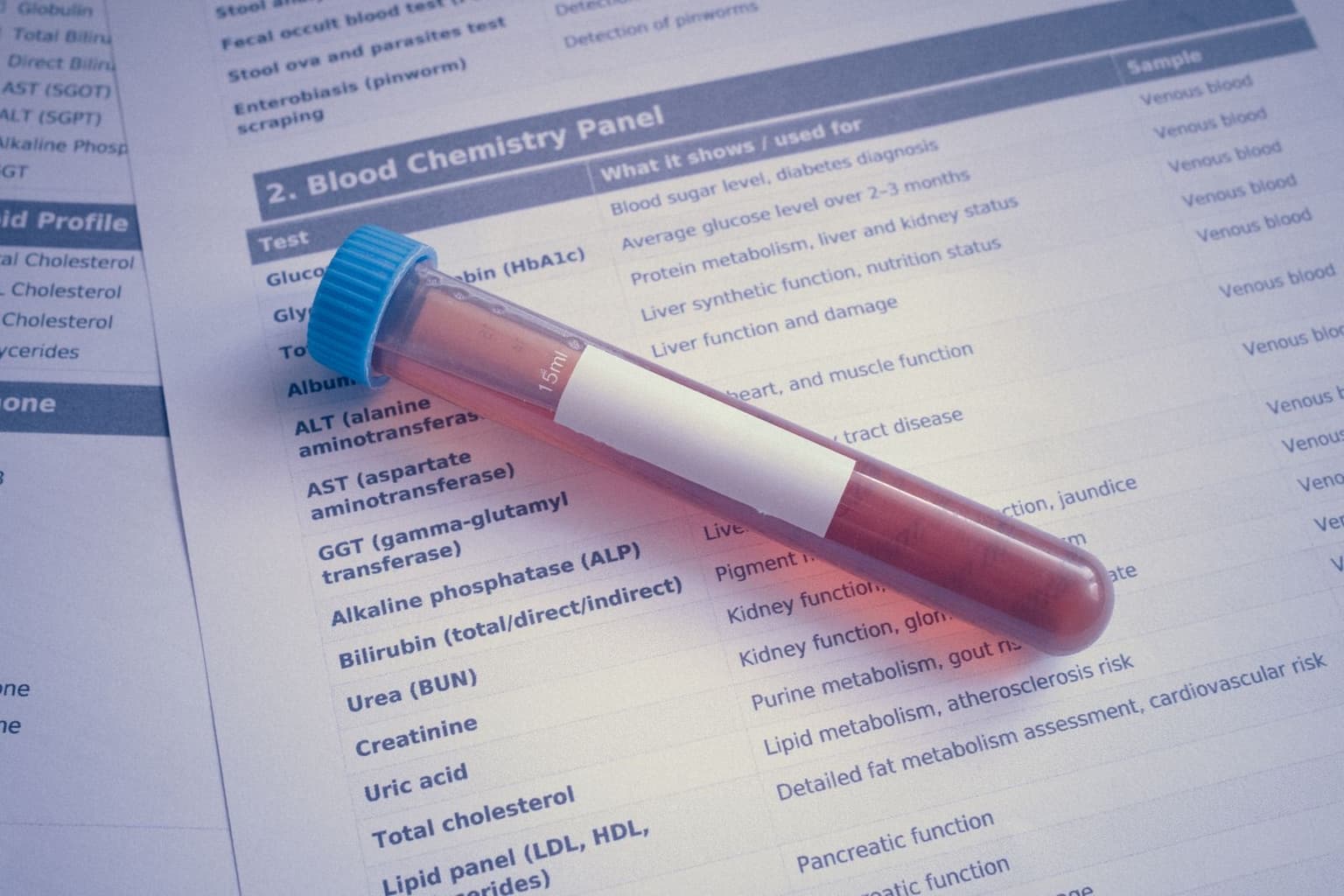
Routine blood work for autoimmune and connective tissue conditions usually includes inflammatory markers. The two most common are the erythrocyte sedimentation rate (ESR) and C-reactive protein (CRP). Both indicate that inflammation is happening somewhere, but CRP responds quickly and directly to inflammatory stimuli, while ESR is only a surrogate marker of the acute phase response.1
- Erythrocyte sedimentation rate (ESR): measures how fast red blood cells settle in a tube over an hour, reported in millimetres per hour. Raised plasma proteins, particularly fibrinogen, make cells clump and fall faster.
- C-reactive protein (CRP): measures the concentration of the protein itself, reported in mg/dL or mg/L. It is made by the liver in response to inflammatory signalling, mainly interleukin-6.
- Neither is specific: a raised result confirms systemic activity but never identifies the organ, joint or disease driving it.
Because both are non-specific, clinicians treat them as one piece of a larger picture rather than a diagnosis. A raised number does not say whether the activity is in your joints, skin or blood vessels, which is exactly why matching lab values against daily functional impact matters.
Why do ESR and CRP respond on different timelines?
Both track systemic inflammation, but on very different biological clocks. CRP has a doubling and decay time of roughly six hours, reaches maximum concentration in under two days, and falls quickly once inflammation resolves.2 ESR depends on the slow accumulation of large plasma proteins, so it takes far longer to reflect a clinical shift.
| Parameter | C-reactive protein (CRP) | Erythrocyte sedimentation rate (ESR) |
|---|---|---|
| Source and mechanism | Made by the liver after interleukin-6 activation | Indirect measure of red cell settling, influenced by plasma fibrinogen |
| Response onset | Synthesis begins roughly 6 to 8 hours after the trigger, peaking at 36 to 50 hours | Rises slowly over days as protein production changes blood density |
| Clearance | Short half-life of about 19 hours | Normalises gradually over weeks after inflammation resolves |
That mismatch explains why blood taken in the first hours of a severe crash can show a sharp CRP spike next to a completely normal ESR. As a flare subsides the reverse happens: CRP drops quickly while ESR stays raised for weeks, reflecting past tissue stress rather than current disease activity.
What do the numbers mean, and why do they disagree?
Reference ranges give context without inviting panic. Laboratories set their own cut-offs, but a normal CRP is generally under 0.9 mg/dL, and results over 50 mg/L are associated with acute bacterial infection about 90% of the time.3 Typical ESR ranges run 0 to 15 mm/hr for adult men and 0 to 20 mm/hr for adult women, and baseline values rise naturally with age.
- Raised CRP with normal ESR: often caught early in an acute flare, after a joint injury, or during active infection, because CRP is made so quickly.
- Raised ESR with normal CRP: commonly seen in chronic low-grade autoimmune activity, kidney impairment, anaemia, or during recovery when fibrinogen is still high.
- Genuine discordance: in a hospital study of patients who had both measured on the same day, a small minority had clearly discordant results, and infection, renal insufficiency and low serum albumin were the factors most associated with a high ESR paired with a low CRP.4
Discordance between your two results is a recognised phenomenon, not a laboratory error and not a sign that your symptoms are imagined. Knowing that markers can diverge is often what makes a reassuring-looking blood draw easier to sit with. The same holds when every test comes back normal and you still feel ill.
What do raised markers signal in rheumatoid arthritis?
In rheumatoid arthritis, rheumatologists use ESR and CRP to monitor synovial inflammation and assess structural risk. Sustained elevations reflect active joint lining inflammation, and markers measured early in the disease are among the laboratory predictors of later joint damage in the hands and feet.
Domain-based tracking for joint disease
To know whether a lab spike matches genuine disease activity, separate daily symptoms by domain instead of flattening them into one pain score. The arthritis support guide covers this in more detail.
- Joint pain by location: which specific joints, wrists, knuckles or knees, were affected during daily tasks.
- Morning stiffness in minutes: how long after waking before tightness eases, timed rather than estimated.
- Swelling and mobility: visible puffiness plus functional limits such as difficulty opening jars or holding cutlery.
- Symmetry: whether both sides are involved, mapped against the dates of blood draws.
How are inflammation markers read in lupus?
Systemic lupus erythematosus behaves differently. Compared with rheumatoid arthritis, people with lupus are more likely to show a raised ESR than a raised CRP, and CRP tends not to track other markers of lupus disease activity such as anti-double-stranded DNA antibodies and complement levels.5
- Separate the domains: record fatigue, joint stiffness and skin changes individually to capture multi-system activity.
- Log environmental triggers: UV exposure, heat and significant stress, recorded next to medication doses.
- Watch for infection: a sudden CRP surge in someone with lupus often signals a secondary bacterial infection rather than a lupus flare on its own.
Recording sun exposure and doses alongside routine bloods gives your rheumatologist the context to tell a classic flare from an infection needing antibiotics. The method is the same one used for tracking lupus triggers across UV, stress and weather.
How do you handle the anxiety of fluctuating results?
Waiting on results is hard, particularly when blood work comes back normal during a bad crash or unexpectedly high on a stable week. Those swings are a limitation of single-point-in-time testing rather than a verdict on how you feel.
Short appointments and repeated retelling make self-advocacy exhausting. Looking at patterns across weeks and months gives a truer picture of your health than any isolated draw, and it is a far lighter thing to carry than trying to justify each individual number.
How do you track symptoms alongside blood work?
The aim is a record your clinician can read in under a minute, next to the lab report. A symptom tracker worksheet is enough to start, and the entries that carry the most weight are the concrete ones.
- Log close to the moment: short entries made on the day beat detailed ones reconstructed a fortnight later.
- Note the date of every draw: so symptoms can be lined up against the result rather than remembered around it.
- Keep functional detail: minutes of morning stiffness, tasks stopped, rest needed.
- Review across weeks: single days mislead, repeated patterns do not.
Bringing that alongside your results turns a confusing number into a trend a specialist can act on.
Frequently asked questions
What causes high ESR and CRP together?
Both rise with inflammation, so they are often raised together by an infection, an autoimmune or inflammatory condition such as rheumatoid arthritis or lupus, an injury or recent surgery, and sometimes cancer. Neither test says which, so your doctor reads them alongside your symptoms, an examination and other tests.
Should I be worried about high ESR and CRP?
A raised result is a reason to follow up, not a diagnosis. Your doctor will look at how high it is, whether it is changing and what else is going on. Get medical advice promptly if raised markers come with a fever, night sweats, unexplained weight loss or feeling very unwell.
What is the normal range for a CRP blood test?
A normal CRP level is generally less than 0.9 mg/dL. Laboratories often use different reference ranges, so it is worth reviewing your specific result with your doctor. CRP is an acute-phase reactant, which means it rises rapidly in response to inflammation.
Why would my ESR be high but my CRP normal?
Discordant results are common: one hospital cohort found the two tests disagreed in 12.5% of patients. ESR is a slower marker that can stay raised for weeks, while CRP drops quickly. Discordance is also frequently seen in systemic lupus erythematosus.
Can I have a flare if my inflammation markers are normal?
Yes. Many people have severe symptoms despite normal ESR and CRP levels. This is why it helps to track functional impact, such as morning stiffness duration in minutes or the location of joint pain, alongside lab work so your doctor sees the complete picture.
How fast does CRP rise during an inflammation spike?
C-reactive protein can rise within about 6 to 10 hours of tissue injury or an inflammatory stimulus, reaching maximum concentration in under two days. That makes it a much faster indicator of acute inflammation than an ESR test.
Does a high CRP always mean a chronic illness flare?
No. A high CRP signals that inflammation is present somewhere in the body, but it does not specify the cause, which can range from an infection to a recent injury. It needs to be read alongside your symptoms and history.
Tools that go with this article
Sources
- https://australianprescriber.tg.org.au/articles/erythrocyte-sedimentation-rate-and-c-reactive-protein.html
- https://pmc.ncbi.nlm.nih.gov/articles/PMC3826426/
- https://my.clevelandclinic.org/health/diagnostics/23056-c-reactive-protein-crp-test
- https://www.ncbi.nlm.nih.gov/books/NBK519570/
- https://pmc.ncbi.nlm.nih.gov/articles/PMC5938146/
Turn day-to-day notes into a clearer health story
Juno helps you track symptoms through conversation, notice patterns, and prepare a structured summary for appointments. See how Juno works. Download Juno on the App Store.
This article is for general information only and does not replace advice from your own clinician. Juno is a health assistant, not a medical provider.
Last reviewed: 2026-09-02